Medical/Ring-Shaped Ultrasound Imaging System
Medical imaging such as X-ray CT, MRI, and ultrasound is essential for diagnosing diseases throughout the body, however, there has been limitation for intracranial imaging in children. If pediatric intracranial imaging becomes possible, a variety of clinical applications can be expected, including the evaluation of brain damage caused by falls, which are common in children, and the evaluation of brain damage associated with neonatal asphyxia. We focus on ultrasound CT as a method to achieve intracranial ultrasound imaging. Ultrasonic CT is a tomographic imaging method proposed by Greenleaf belonged to Mayo Clinic in the 1970s that uses a ring array consisting of approximately 2000 elements. At that time it was difficult to solving complex ultrasound propagation paths with computers, and therefore it was not in practical use. The purpose of this research is to develop ultrasound CT technology that enables pediatric intracranial imaging. We will develop an imaging algorithm using a prototype ultrasound CT system, a skull phantom, a skull specimen, etc., as well as extract target diseases for pediatric intracranial imaging, and examine performance indicators required for diagnostic images.
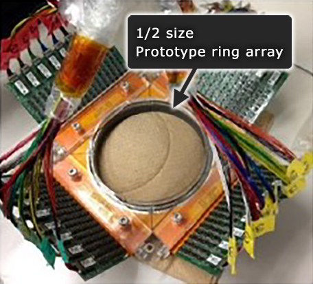
【Prototype ultrasonic CT device】
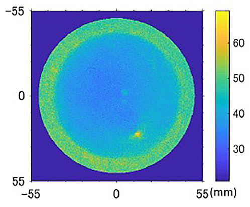
【Imaging results using a skull phantom】
共同研究者
東京医科歯科大学(藤岡友之氏)、超音波CT装置メーカ:㈱Lily MedTech(東隆氏)、東北大学(荒川元孝氏)、都立大学(関根紀夫氏、伊藤恵理子氏)